
Figure 2.
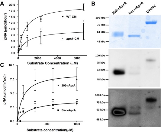
AprA has DPPIV‐like enzymatic activity. (A) Wild‐type or aprA− conditioned media were incubated with various concentrations of H‐Gly‐Pro‐pNA p‐tosylate and the formation of the pNA digestion product was measured. (B) 293‐rAprA, Bac‐rAprA, and rDPPIV were electrophoresed on SDS‐polyacrylamide gels. The upper panel shows Coomassie blue staining of a gel; the middle and lower panels show a corresponding Western blot stained with biotinylated Lens culinaris agglutinin. (C) 293‐rAprA and Bac‐rAprA were incubated with various concentrations of H‐Gly‐Pro‐pNA p‐tosylate as in panel A. Values in A and C are mean ± SEM, n = 3 (n = 7 for Bac‐rAprA in C). Asterisk indicates P < 0.05, **P < 0.01 (t test).