Fig. 1.
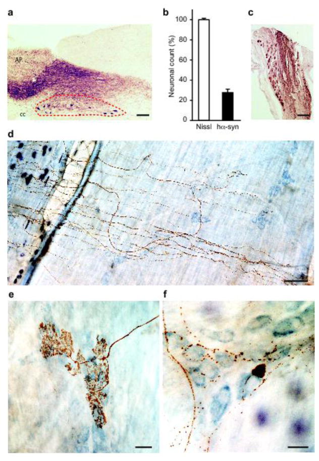
Accumulation of hα-synuclein in the DMnX, nodose ganglion and gastric wall after injections of hα-synuclein-AAVs into the vagus nerve. a–c Rats (n = 5) received a single injection of hα-synuclein-carrying AAVs into the left vagus nerve. Analyses were performed at 2–3 weeks post-treatment. a A representative image shows a section of the medulla oblongata immunostained with anti-hα-synuclein; the left DMnX is delineated by dashed lines, and the area postrema (AP) and central canal (cc) are indicated. Scale bar = 100 μm. b Medulla oblongata sections were used for stereological counting of Nissl-stained neurons (empty bar) and hα-synuclein-immunoreactive cells (solid bar) in the left DMnX. Values (means ± SEM) are expressed as percent of the total number of Nissl-stained neurons. c The representative image shows neurons robustly labeled with anti-hα-synuclein in a section of the left nodose ganglion. Scale bar = 100 μm. d–f Rats (n = 5) were killed 6 to 12 months after a single injection of hα-synuclein-carrying AAVs into the left vagus nerve. Stomach whole mounts were stained with anti-hα-synuclein and counterstained with Cuprolinic Blue. Representative images show immunoreactive fibers and nerve terminals: long intramuscular arrays (IMAs) of rectilinear terminals (d), a single vagal afferent terminating as highly arborizing intraganglionic laminar endings (IGLEs) (e), and varicosity-rich fibers with morphological features of preganglionic vagal efferents (f). Scale bars = 100 μm in (d), 25 μm in (e) and 20 μm in (f).

