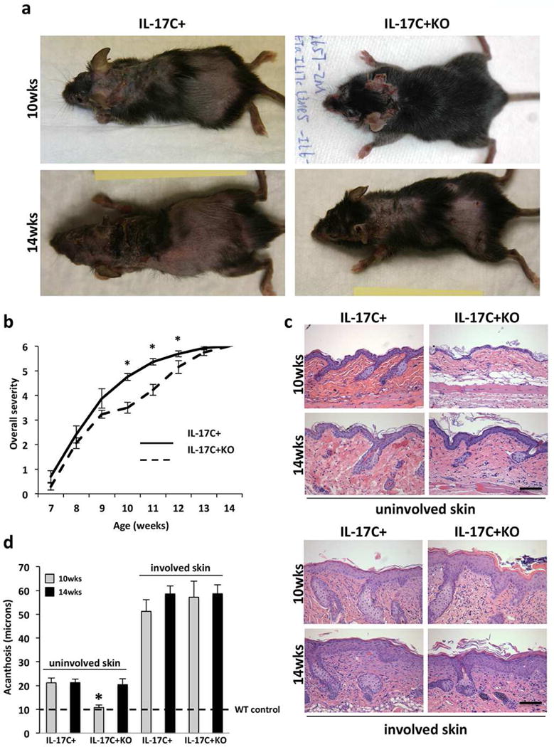
Figure 1. IL-17C+KO mice develop a delayed inflammatory skin phenotype.

(a) Representative images of IL-17C+ and IL-17C+KO mice at 10- and 14-weeks of age. (b) Mouse overall severity scale between 7- and 14-weeks of age. (c) Representative images of H&E stained uninvolved and involved skin from IL-17C+ and IL-17C+KO mice at 10- and 14-weeks of age. (d) Mean epidermal thickness (acanthosis; μm) measures (± SEM) of involved and uninvolved dorsal skin of IL-17C+ (n=8 at 10 weeks; n=5 at 14 weeks) and IL-17C+KO (n=8 at 10 weeks, n=8 at 14 weeks) mice at 10-weeks and 14-weeks of age. The dashed line indicates wildtype mouse levels. Scale bars in c = 100 μm; *P<0.05.
