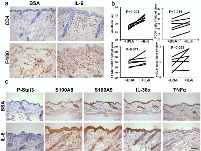
Figure 3. Intradermal IL-6 injection into IL-17C+KO mouse skin elicits a IL-17C+ skin phenotype.

(a) CD4+ and F4/80+ immunohistochemistry on IL-17C+KO mice skin intradermally injected with BSA or recombinant IL-6 every other day for 16 days. (b) Quantification of epidermal thickness (acanthosis, μm) and infiltrating immune cells (CD4+, CD8+ and F4/80+; mean number of cells/field of view) in IL-6 and BSA injected IL-17C+KO (n=6). Significance values are as indicated (b) Representative images from IL-17C+KO skin injected with either BSA or IL-6 immunostained for phospho-Stat3, S100A8/A9, IL-1F6 and TNFα. Scale bar in a and c = 100 μm.
