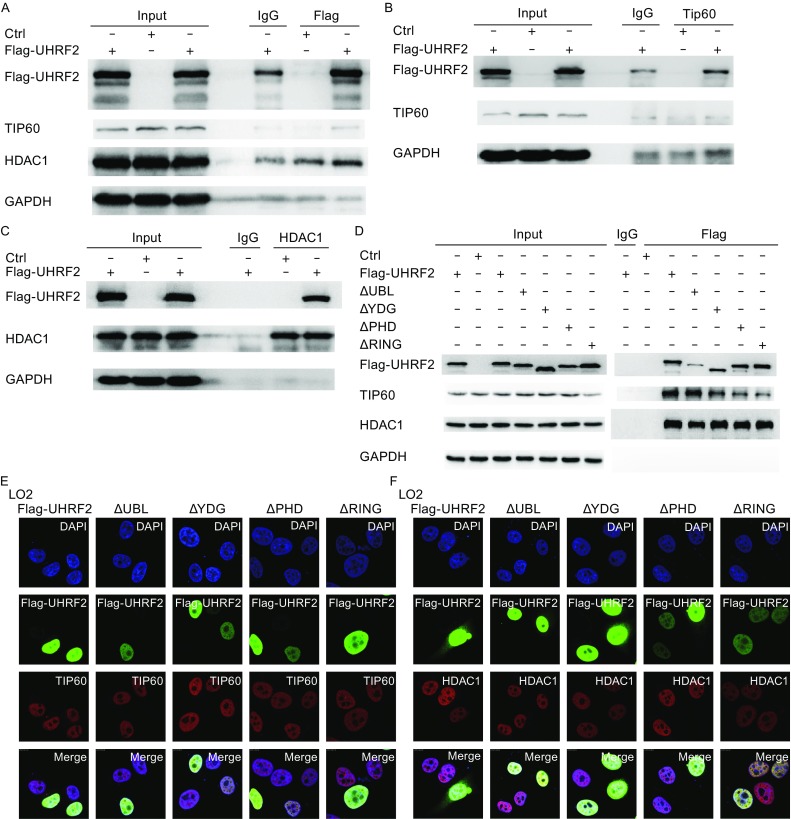
Figure 2.

UHRF2 interacts with TIP60 and HDAC1. (A) HEK293 cells were transfected with control or Flag-UHRF2 plasmids, respectively. After 48 h, the total cellular lysates were immunoprecipitated using anti-Flag antibodies. (B) HEK293 cells were transfected with plasmids as shown. Cellular lysates were immunoprecipitated with anti-TIP60 antibodies and analyzed by Western blot. (C) HEK293 cells were transfected with the indicated plasmids. Total cellular lysates were subjected to HDAC1 immunoprecipitation. (D) HEK293 cells were transfected with full-length UHRF2 or various deletion mutant constructs of UHRF2 plasmids. Cellular lysates were immunoprecipitated using anti-Flag antibodies and analyzed by Western blot. (E and F) Double-labeling immunofluorescence combined with CLSM observation revealed co-localization of TIP60 and UHRF2 in LO2 cells. Co-localization of HDAC1 and UHRF2 was also revealed along with nuclear expression of all proteins
