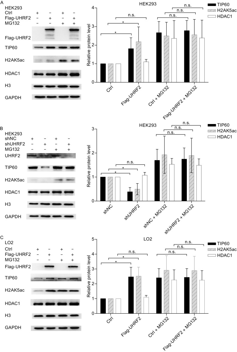
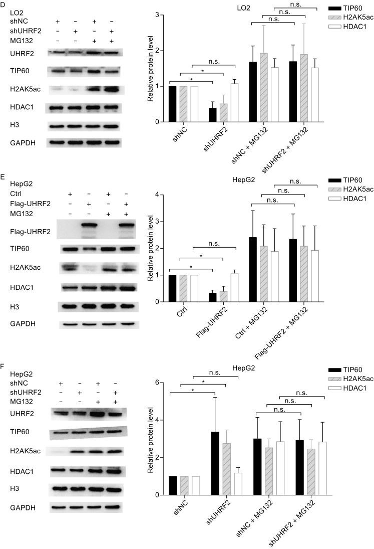
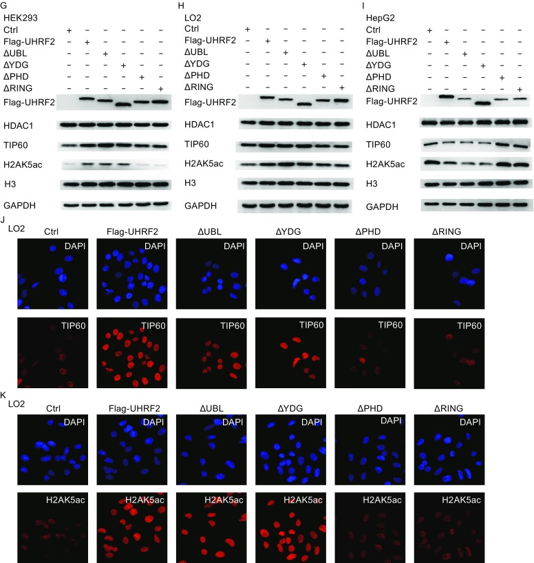
Figure 3.


UHRF2 regulates the expression and activity of TIP60. (A, C and E) HEK293, LO2 and HepG2 cells were transfected with control or Flag-UHRF2 plasmids. They were exposed to MG132 (10 μg/mL) for 12 h and harvested. The cellular lysates were analyzed by Western blot. (B, D and F) HEK293, LO2 and HepG2 cells were transfected with shNC or shUHRF2. After 48 h, the total cellular lysates were analyzed by Western blot. Data were expressed as means ± SD, n = 3. Significance was indicated as *P < 0.05, and non-significance as n.s P > 0.05. (G–I) HEK293, LO2 and HepG2 cells were transfected with the plasmids as shown. The cellular lysates were analyzed by Western blot. (J and K) Immunofluorescence analyses were performed using anti-TIP60 or anti-HDAC1 antibodies
