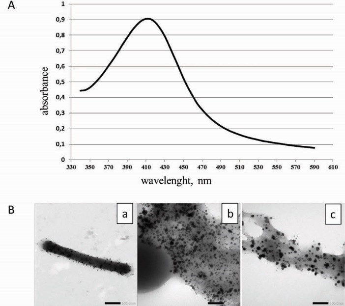
Fig. 1.

A- UV/VIS absorption spectra of the Ag NPs prepared using L. plantarum 92T cell suspension. B- Transmission of electron micrographs of Ag NPs obtained with lactobacilli after 24 h of incubation in AgNO3 solution. (a) – cell of strain L. fermentum 215 with Ag NPs on the surface (scale bar corresponds to 500 nm); (b, c) – accumulation of Ag NPs in capsular polymers of L. casei strain (scale bar corresponds to 100 nm).
