
Figure 2.
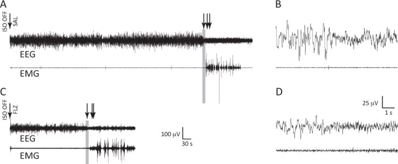
EEG/EMG recordings of the transition to arousal from anesthesia. A, Representative waveforms from an animal given saline (SAL) via tail vein catheter at the cessation of isoflurane anesthesia. EEG from the medial (hippocampal) lead is shown. Arrows designate (from left to right): isoflurane cessation and administration of saline, EEG wake (defined as a transition to lower amplitude, faster oscillatory activity), eye blink, and ambulation. B, Time-expanded 10-second epoch capturing the transition to a wakeful EEG (before the onset of movement). The gray rectangle in A corresponds to this expansion in B. C, Representative waveforms from an animal given flumazenil (FLZ) via tail vein catheter at the cessation of isoflurane anesthesia. Arrows designate (from left to right): isoflurane cessation and administration of flumazenil, EEG wake, eye blink, and ambulation. D, Time-expanded 10-second epoch capturing the transition to a wakeful EEG (before the onset of movement). The gray rectangle in C corresponds to this expansion in D.