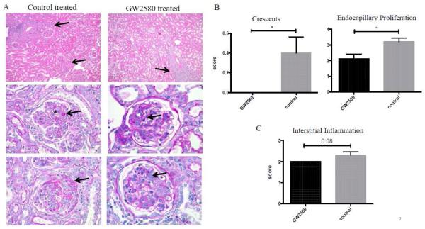
Fig. 2.
Renal histopathology. The left side of panel A shows representative images of control treated mice with interstitial inflammation (arrows, top left), deposits (arrow) with endocapillary proliferation (*) (middle left), and necrotizing crescent (arrow, bottom left). GW2580 treated mice displayed improved histopathology, although still displayed patchy interstitial inflammation (arrow, top right), mesangial deposits (arrow, middle right), and less extensive endocapillary proliferation (arrow, bottom right). Scores for crescents, endocapillary proliferation, and interstitial inflammation are shown in B and C. (GW2580 treated, n=9; Control treated, n=10) (*p<0.05).

