
FIGURE 4.
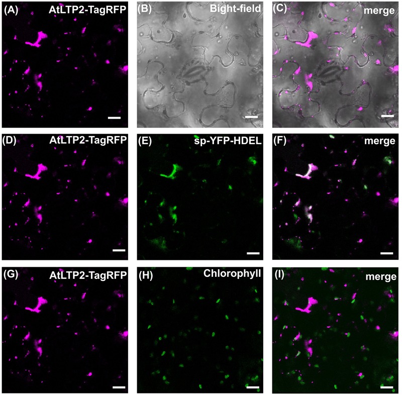
The secretory pathway is involved in both cell wall and plastids AtLTP2-TagRFP localization. The AtLTP2-TagRFP fusion protein was produced together with the endoplasmic reticulum (ER) marker sp-YFP-HDEL in N. benthamiana leaves which were subjected to Brefeldin A (BFA) treatment. The AtLTP2-TagRFP fluorescence signal is false-colored in magenta in (A,D,G). The bright-field (B) is displayed to visualize the cell morphology. ER marker (E) and chlorophyll auto-fluorescence (H) are false-colored in green and the merge is presented in white in the right column (C,F,I). Note that in presence of BFA, AtLTP2-TagRFP fluorescence is restricted to the ER-Golgi hybrid and BFA compartments. Scale bars: 40 μm.