FIGURE 8.

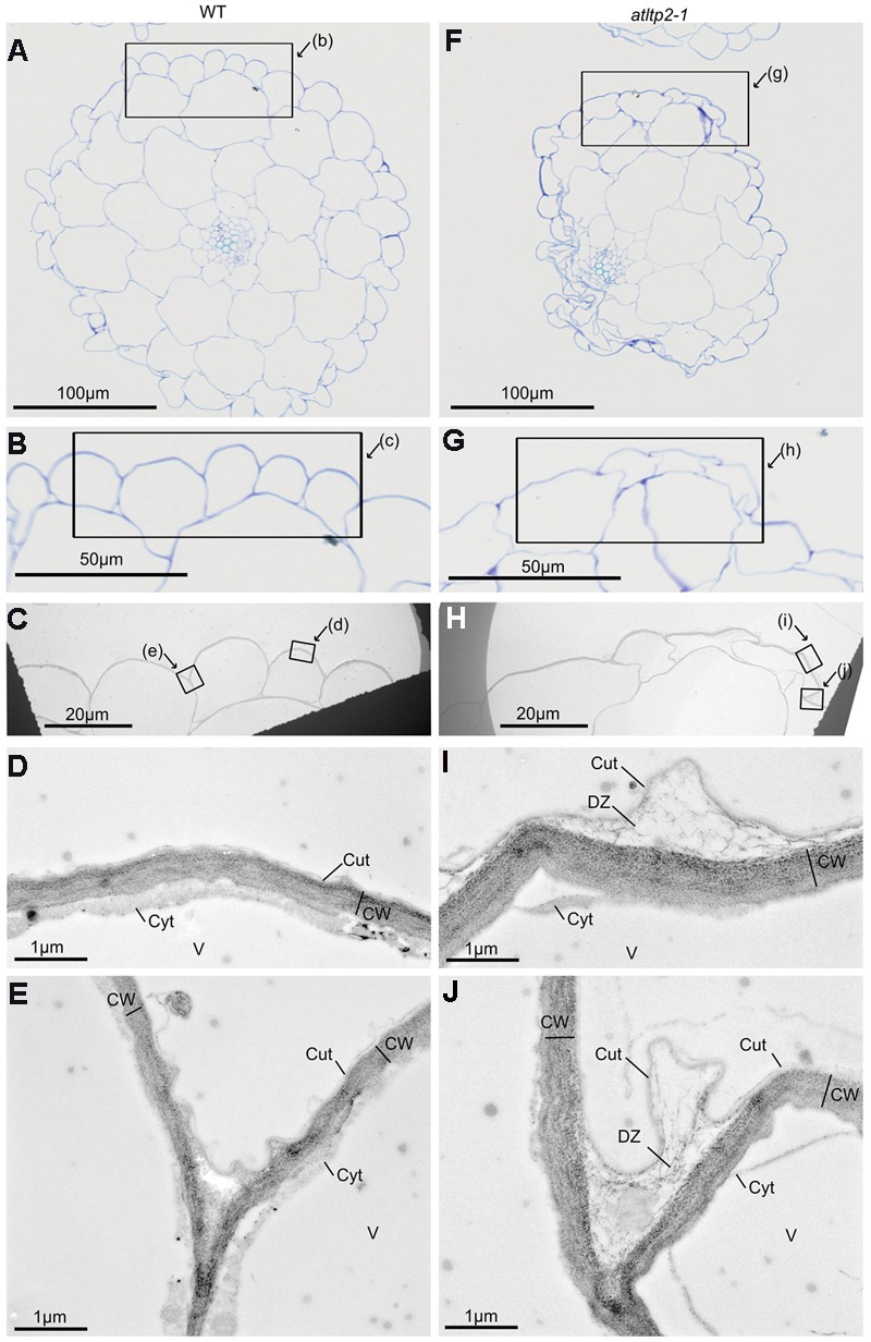
Correlated light microscopy (LM) and TEM observations of 5-day-old etiolated hypocotyls of WT and atltp2-1. Semi-thin cross-sections of WT (A–E) and of atltp2-1 (F–J) were stained with TB for LM observations (A,B,F,G) and ultra-thin serial sections were PATAg-labeled for TEM observations (C–E,H–J). The frames indicate the magnified zones. Note the overall shrinkage of atltp2-1 hypocotyl, although no particular phenotype was observable at the cuticle-cell wall interface at the correlated high magnification of LM (B,G) and low magnification of TEM (C,H), respectively. In the atltp2-1, higher TEM magnification (I,J) allowed, the visualization of the PATAg-reactive cell wall defibrillated material at the detachment zone located between the cuticle and the cell wall. Legend: cuticle (Cut); cytoplasm (Cyt); cell wall (CW); cuticle-cell wall detachment zone (DZ); vacuole (V).
