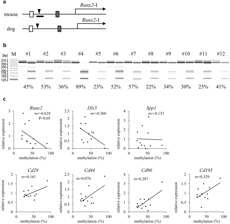
Fig. 4.
Correlation between DNA methylation and gene expression among primary cultures of canine marrow (from different dogs). a) Structure of the proximal Runx2 promoter in the mouse genome and dog genome is shown. White and shaded boxes indicate conserved regions corresponding to each color. Arrowheads mark mouse CpG-2,505 and canine CpG-2,829. The range of defined Runx2-DMR is indicated as a horizontal bar in the mouse panel. b) A gel image as output from the results of combined bisulfite restriction analysis (CoBRA) targeting CpG-2,829 in canine marrow cultures. Fragments digested with HpyCH4IV are loaded onto odd lanes, except for the leftmost lane (showing a 25-bp DNA ladder; M). In the lanes immediately on the left, the fragment without the digestion is loaded as the experimental control. Each number on the lanes is assigned to each individual. The PCR product 315 bp long is digested into fragments 128 and 187 bp long when canine CpG-2,829 is methylated. Various rates of methylation are shown as indicating each score below their lanes. c) Marrow cells derived from each individual are plotted on the scatter diagrams. Vertical and horizontal scales indicate relative expression levels of genes headlining each diagram to the sample mean and the methylation rate of canine CpG-2,829, respectively. Statistical significance of correlation is shown only between CpG-2,829 methylation and Runx2 expression.

