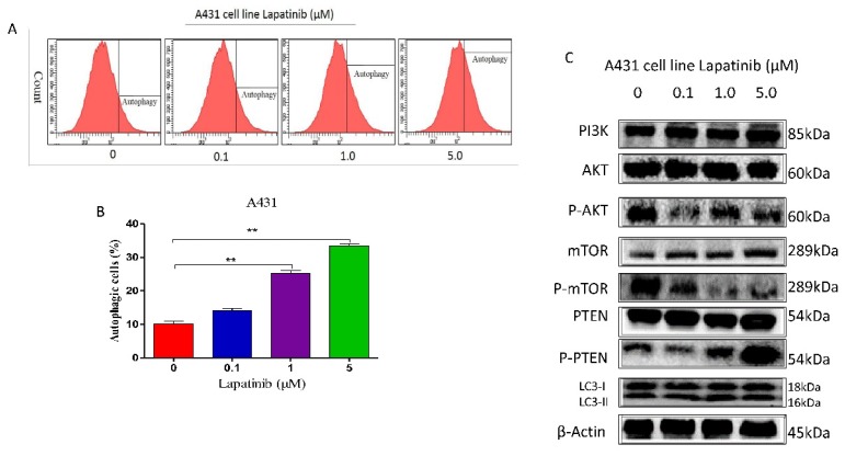
Figure 6.
A. The cellular autophagy induced by the concentration of lapatinib (0-5µM) by confocal microscopy with the application of Cyto-ID autophagy detection kit. B. The percentage of autophagic A431 cells was stained by the Cyto-ID autophagy detection kit and analyzed using the green (FL1) channel of the flow cytometer. C. The protein markers as shown on figure were detected by western blot assay.

