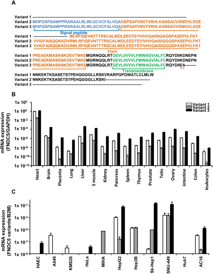
Figure 1. Schematic sequence of human FNDC5 variants and mRNA expression of FNDC5 variants in human tissues and cell lines.
(A) Multiple amino acid sequence alignment of the variants of the human FNDC5 gene. Variant 1 contains no signal peptide and is cleaved to the irisin sequence. Variants 2 and 3 have a different start codon (ATA) than variant 1, and the C-terminal amino acids are KD → EA in variant 3 (blue = signal peptide, orange = irisin, green = transmembrane sequence). (B) The mRNA levels of the three FNDC5 variants in human tissues are shown. (C) The mRNA levels of the three FNDC5 variants are shown for human cell lines. The expression level of each sample was measured by quantitative real-time RT-PCR using GAPDH or B2M for normalization. HAEC; human aortic endothelial cell, A549; adenocarcinomic human alveolar basal epithelial cells, KMS26; Plasma cell myeloma cell, HeLa; cervical adenocarcinoma cell line, MIHA; nontumorigenic immortalized human hepatocyte cell line, HepG2, Hep3B, Sk-Hep1, SNU449, and Huh7: human hepatoma cell lines, AC16: human cardiomyocyte cell line.

