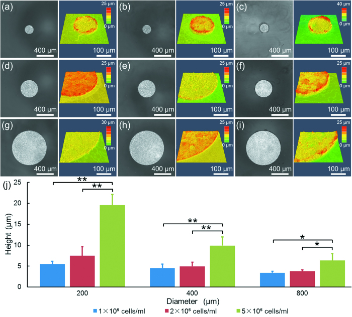
Figure 3.
The microscopic and corresponding confocal images of A549 cell clusters as a function of initial diameters and seeding densities at 200 μm and 1 × 10⁵ cells/ml (a), 200 μm and 2 × 10⁵ cells/ml (b), 200 μm and 5 × 10⁵ cells/ml (c), 400 μm and 1 × 10⁵ cells/ml (d), 400 μm and 2 × 10⁵ cells/ml (e), 400 μm and 5 × 10⁵ cells/ml (f), 800 μm and 1 × 10⁵ cells/ml (g), 800 μm and 2 × 10⁵ cells/ml (h), 800 μm and 5 × 10⁵ cells/ml (i). The quantified heights of cell clusters were summarized in (j) (*p < 0.05, **p < 0.01).

