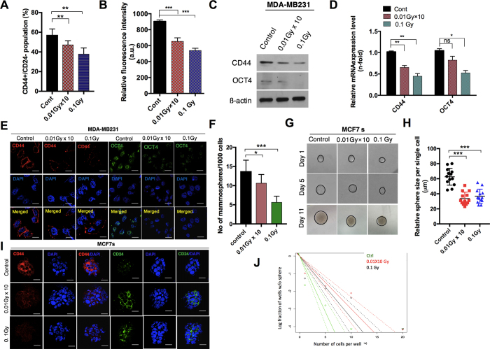
Figure 1. Low-dose radiation decreases the cancer stem-cell maintenance in breast cancer cell lines.
(A) Flow cytometer analysis of CD44+/CD24− cells in control and LDR-exposed MDA-MB231 cells at 0.01 Gy × 10 (fractionated) and 0.1 Gy (single dose). (B) Determination of the CD44 fluorescence intensity in LDR treated and untreated control cells using an Elisa reader. (C) Western blot of the CD44 and OCT4 protein levels LDR treated and untreated control MDA-MB231 cells. (D) qRT-PCR analyses results of CD44 and OCT4 gene expression levels in LDR treated and untreated control MDA-MB231 cells. (E) Immunocytochemistry of CD24 and OCT4 expression levels in LDR treated and untreated control cells. (F) Determination of the sphere-forming ability of LDR-exposed and control MCF7 breast cancer cells cultured in a sphere-conditioned medium. (G,H) Single-cell assay of LDR-exposed and control MCF7 sphere cells as observed from days 1 to 11 days after treatment. Quantification of the average size of each single cell is shown in the representative graph. (I) Immunocytochemistry of CD24 and CD24 in LDR treated and untreated control MCF7 sphere-cultured cells. (J) Limiting dilution assay performed on MCF7 cells after LDR exposure and compared with LDR unexposed control cells. Solid lines represents the average value of samples. β-actin was used as a loading control. Error bars denote the mean ± S.D. of triplicate samples. *p < 0.05, **p < 0.01, and ***p < 0.001.

