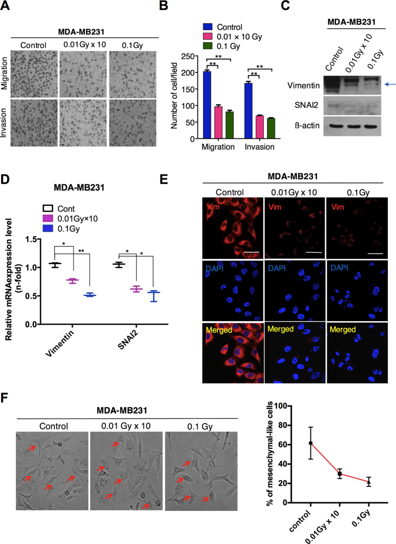
Figure 2. Low-dose radiation reduces the migration and invasion in MDA-MB231 cells via EMT.
(A) Migration and invasion Transwell assays of MDA-MB231 breast cancer cells after LDR at doses of 0.01 Gy × 10 (fractionated) and 0.1 Gy (single dose). (B) Representative graphs of the migration and invasion of LDR-exposed MDA-MB231 cells. (C) Western blot for vimentin and SNAI2 (SLUG) in MDA-MB231 breast cancer cells after irradiation at 0.01Gy × 10 and 0.1Gy. (D) qRT-PCR analysis of vimentin and SNAI2 gene expression after irradiation in MDA-MB231 cells. (E) Immunocytochemistry for a vimentin mesenchymal marker in MDA-MB231 breast cancer cells after irradiation. (F) Phase-contrast images of control and LDR-treated MDA-MB231 cells. Representative graph shows the percentage of mesenchymal-like cells in respective each group. β-actin was used as a loading control. Error bars denote the mean ± S.D. of triplicate samples. *p < 0.05, and **p < 0.01.

