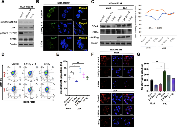
Figure 3. Low-dose radiation inhibits JAK1/STAT3 signaling in breast cancer cells.
(A) Western blot analysis for the phosphorylation status of the JAK1/STAT3 pathway in LDR-exposed MDA-MB231 cells. (B) Immunofluorescence of the pJAK1 expression status in LDR-treated MDA-MB231 cells at similar doses. (C) Western blot analyses of the protein levels of CD24 and CD44 in LDR-irradiated MDA-MB231 cells after JAK1 overexpression. Representative graphs show the intensity of the CD44/CD24 levels in the LDR-exposed MDA-MB231 cells. (D) Flow cytometer analysis of CD44+CD24− cells in control and JAK1-overexpressing LDR-exposed MDA-MB231 cells at 0.01Gy × 10 (fractionated) and 0.1Gy (single dose). (E) Representative graph of the CD44+CD24− population in Mock and JAK1-overexpressing LDR-irradiated cells at similar doses. (F) Immunofluorescence staining of the CD44 expression outcomes in LDR-exposed MDA-MB231 cells after JAK1 overexpression. (G) Analysis of invasive cells in LDR-irradiated JAK1-overexpressing MDA-MB231 cells. β-actin was used as a loading control. Error bars denote the mean ± S.D. of triplicate samples. *p < 0.05, and **p < 0.01.

