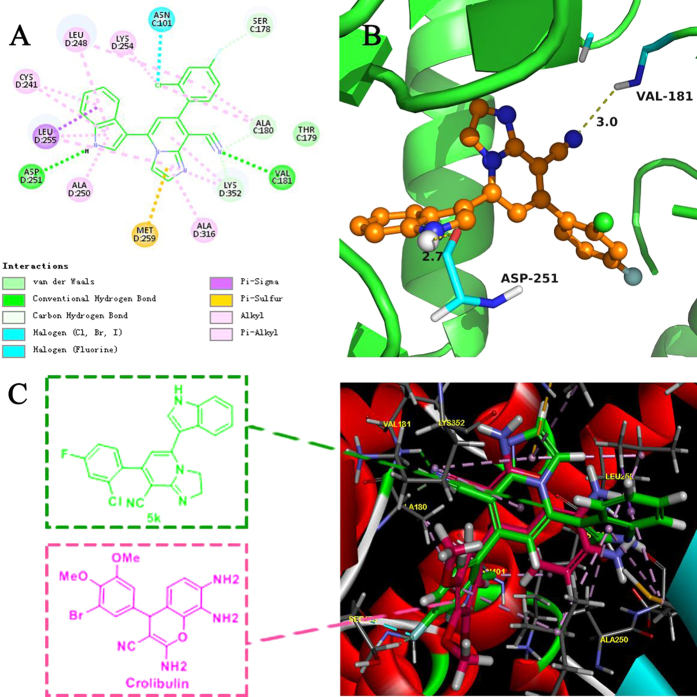
Figure 7. The binding mode between the active conformation of 5k and tubulin.
(A) 2D diagram of the interaction between 5k and the colchicines binding site. (B) 3D diagram of the interaction between 5k and the colchicine binding site. For clarity, only interacting residues are displayed. The H-bond (yellow arrows) is displayed as dotted arrows. (C) Predicted modes for 5k (green) and Crolibulin (pink) binding at the colchicine-binding site of tubulin (PDB code: 4O2B), and overlapping with each other.

