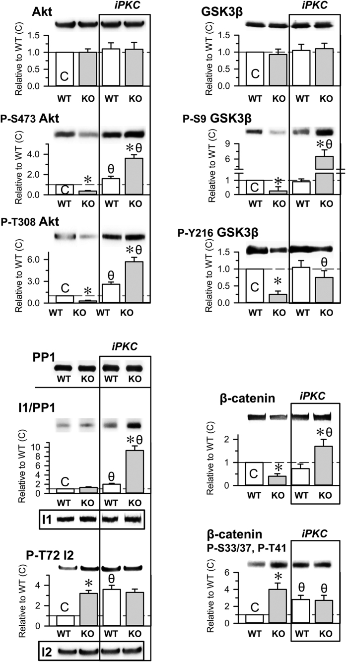
Figure 7. Effects of PKC inhibition on the Akt/GSK3β signaling pathway in HINT1+/+ WT and HINT1−/− KO mice.
The iPKC Gö7874 (1 nmol, icv) was injected 30 min before euthanasia. The levels of the signaling proteins and of their phosphorylated forms were determined in synaptosomes obtained from mouse prefrontal cortices. Representative blots are shown. Tubulin was used as a loading control. Each bar represents the mean ± SEM of the data from at least three determinations, which were performed using different gels and blots. An arbitrary value of 1 was assigned to the control HINT1+/+ WT data (C). *Significant difference between the KO (grey bars) and the WT group (open bars); θ indicates a significant differences caused by Gö7874 but within each group of mice, WT and KO. ANOVA, all pairwise Holm-Sidak multiple comparison tests, p < 0.05.

