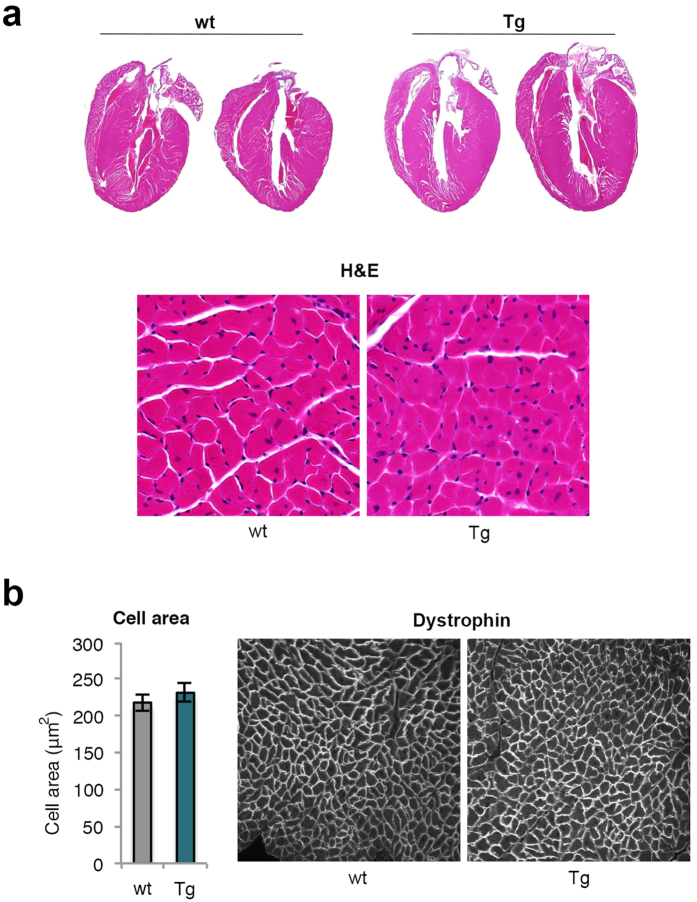
Figure 2. Increased heart size and cell number but not cell size in transgenic mice.
(a) Representative heart sections from 23 weeks old wt and Tg mice on HFD and the left ventricular wall (20x magnification) visualized with Hematoxylin staining. (b) Quantification of cell area (n = 4/group) of the left ventricular wall (20x magnification) stained with anti-dystrophin. The experimental data are presented as means ± SEM. Student’s t-test was used.

