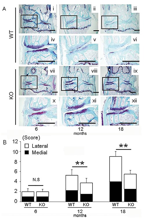
Figure 1.

A SFO staining of knee joints in WT mice (i-iii) and LOX-1 KO mice (vii-ix) at 6, 12, and 18 months. Insets show higher-magnification views of the medial compartment from WT (iv-vi) and LOX-1 KO mice (x-xii). B) The summed OARSI scores of tibias and femurs in WT and LOX-1 KO mice at 6, 12, and 18 months. The white and black coloring represents the scores of lateral and medial compartments. Values shown represent the mean ± SD. **P<0.001. Scale bar: 500 µm.
