
Figure 3.
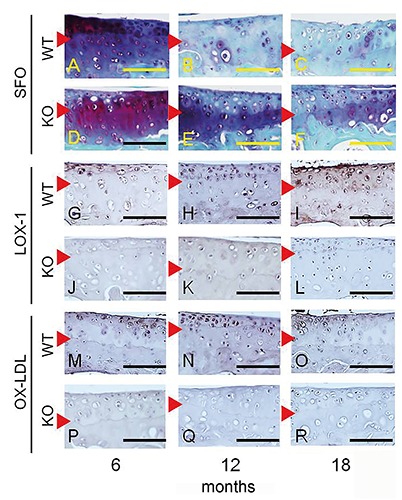
Immunostaining for LOX-1 and ox-LDL expression in the articular cartilage of the medial tibia. Corresponding SFO-stained images for each section (A-F). Positive staining for LOX-1 (G) and ox-LDL (M) in chondrocytes from 6-month-old mice. LOX-1 and ox-LDL staining in chondrocytes increased at 12 months (H, N) and 18 months (I, O) of age. LOX-1 and ox-LDL expression was negative in chondrocytes from LOX-1 KO mice (J-L and P-R). Red arrowhead: tide mark. Scale bar: 100 µm.