Figure 2.

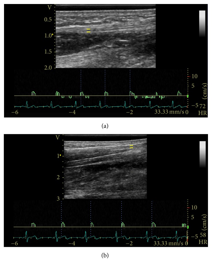
Sample PW Doppler examination in the (a) neutral position and (b) flexed 30° position with spectral tracing of the median nerve proximal to the wrist crease. The yellow bars indicate the gate or sample volume.

Sample PW Doppler examination in the (a) neutral position and (b) flexed 30° position with spectral tracing of the median nerve proximal to the wrist crease. The yellow bars indicate the gate or sample volume.