
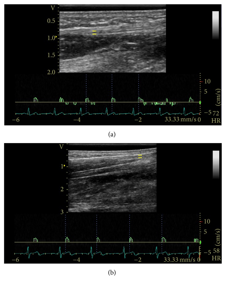
Figure 2.
Sample PW Doppler examination in the (a) neutral position and (b) flexed 30° position with spectral tracing of the median nerve proximal to the wrist crease. The yellow bars indicate the gate or sample volume.
Official websites use .gov
A
.gov website belongs to an official
government organization in the United States.
Secure .gov websites use HTTPS
A lock (
) or https:// means you've safely
connected to the .gov website. Share sensitive
information only on official, secure websites.
Sample PW Doppler examination in the (a) neutral position and (b) flexed 30° position with spectral tracing of the median nerve proximal to the wrist crease. The yellow bars indicate the gate or sample volume.