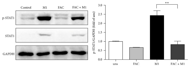
Figure 5.
Effect of iron on STAT1 activation in M1 macrophages. RAW264.7 macrophage cells were incubated with the stimulus, either FAC, IFN-γ, or no stimulus for 24 h. Cell lysates were prepared, and phosphorylation of STAT1 (p-STAT1) was analyzed by western blot. One representative blot and the densitometric quantification are shown. Data are mean ± SD for three independent experiments. (uns: unstimulated macrophages; ∗∗p < 0.01).

