Figure 1.
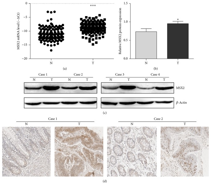
The expression of MSX2 in CRC tumor tissue (T) and adjacent tissue (N). (a) The MSX2 expression of T and N in 136 CRC patients was determined by qRT-PCR (∗∗∗P < 0.001, n = 136). Lower ΔCt value indicates higher MSX2 expression (ΔCt = CtMSX2 − Ctβ-actin). (b) The protein expression of MSX2 of T and N was determined by western blotting (∗P < 0.05, n = 10). The MSX2 expression was normalized to β-actin. (c) Representative western blotting analysis of MSX2 expression in T and N and β-actin was used as a loading control. (d) Representative immunohistochemical analysis of MSX2 expression in T and N.

