Figure 3.
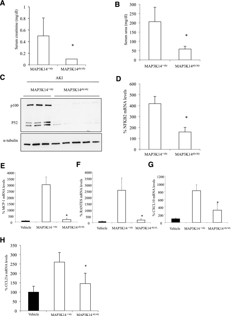
MAP3K14 deficient mice were protected from folate nephropathy–associated AKI. (A) Serum creatinine, *p<0.02 versus heterozygous mice. (B) Serum urea, *p<0.001 versus heterozygous mice. (C) NFκB2 p100 and p52 proteins (representative Western blot). (D) NFκB2 mRNA, *p<0.01 versus heterozygous mice with folate nephropathy. (E) Decreased whole kidney MCP-1, (F) RANTES, and (G) CXCL10 mRNA expression in MAP3K14 deficient mice with folate nephropathy compared with heterozygous mice. *p<0.02 versus heterozygous mice with folate nephropathy. (H) CCL21a mRNA expression. Mean±SD of six mice per group at the 72-hour time-point; *p<0.03 versus heterozygous mice with folate nephropathy.

