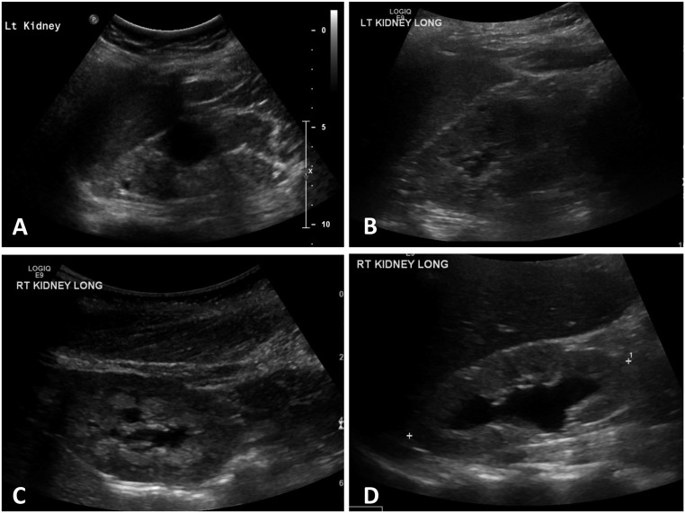
Figure 7.
Renal ultrasonography demonstrates common structural abnormalities associated with Bardet-Biedl syndrome. (A) Demonstrates the typical cystic dysplastic appearance associated with BBS: a small subcortical cyst, one large cyst, and loss of corticomedullary differentiation. (B) Subcapsular cysts and increased echogenicity. (C) Nephrocalcinosis. (D) Renal pelvic dilation. LT, left; RT, right.

