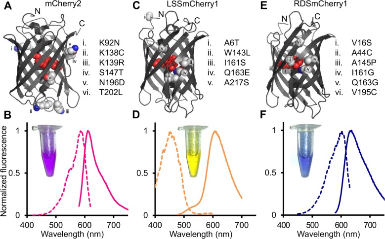
Fig 1. Summary of mutations and fluorescence spectra of mCherry2, LSSmCherry1, and RDSmCherry1.
(A,C,E) Positions of amino acid substitutions mapped onto the crystal structure of mCherry (PDB ID: 2H5Q) [50] for (A) mCherry2, (B) LSSmCherry1, and (C) RDSmCherry1. (B,D,F) Normalized fluorescence excitation (dashed line) and emission spectra (solid line) of (B) mCherry2, (D) LSSmCherry1, and (F) RDSmCherry1. Insets are white light images of purified proteins.

