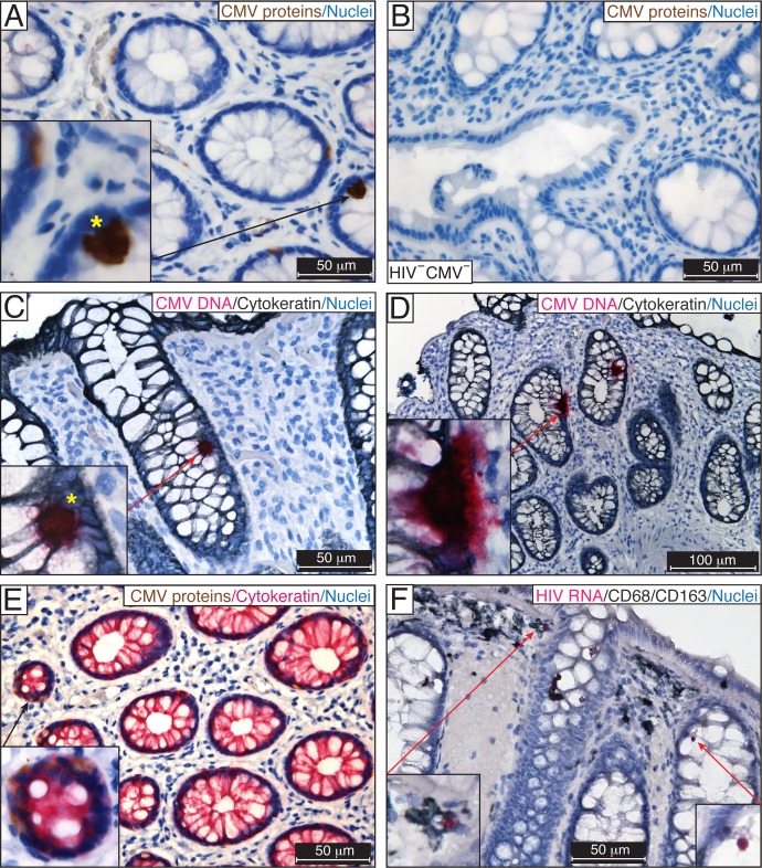
Fig 2. Detection of CMV in rectosigmoid biopsies from ART-suppressed HIV/CMV-coinfected individuals.
IHC for CMV IE, early and late structural proteins using the anti-CMV antibody blend in FFPE rectosigmoid biopsies from ART-suppressed HIV/CMV-coinfected individuals (A) and a HIV-negative, CMV-negative individual (B). The presence of CMV was verified by DNAscope ISH using a probe targeting the noncoding region (120742–122152) of CMV strain Merlin, which corresponds to the UL83 (pp65) sequence (fuchsia) (C, D). Yellow stars indicate similar patterns of IE protein expression (A) and the presence of viral DNA (C) in the nuclei of CMV-infected intestinal cells. (E) CMV early and late proteins (brown) were detected in the cytoplasm of intestinal epithelial cells costained for cytokeratin (fuchsia). (F) HIV RNA was randomly detected by RNAscope ISH in CD68/CD163-positive macrophages (insets) in an adjacent section of tissue shown in (C). Nuclei were counterstained with hematoxylin. Scale bars: 50 μm (A–C, E, F), 100 μm (D). These results are representative of those observed in rectosigmoid biopsies from 9 asymptomatic ART-suppressed HIV/CMV-coinfected individuals.

