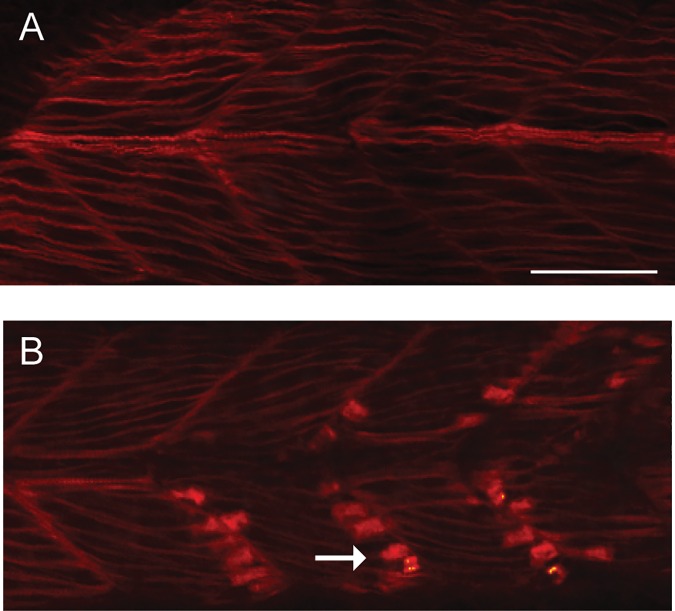
Fig 5. Abnormal phalloidin staining in skeletal muscle from caf mutant embryos.
Wild type (WT) and caf mutants were staining with phalloidin to illuminate filamentous actin and then visualized whole mount by confocal microscopy. (A) WTs show the expected pattern of staining at 24 hpf (n = 5). (B) In muscle from caf mutants, there is an accumulation of intense staining in the region of the myotendinous junction (arrow) (n = 5). Scale bar = 10 um.

