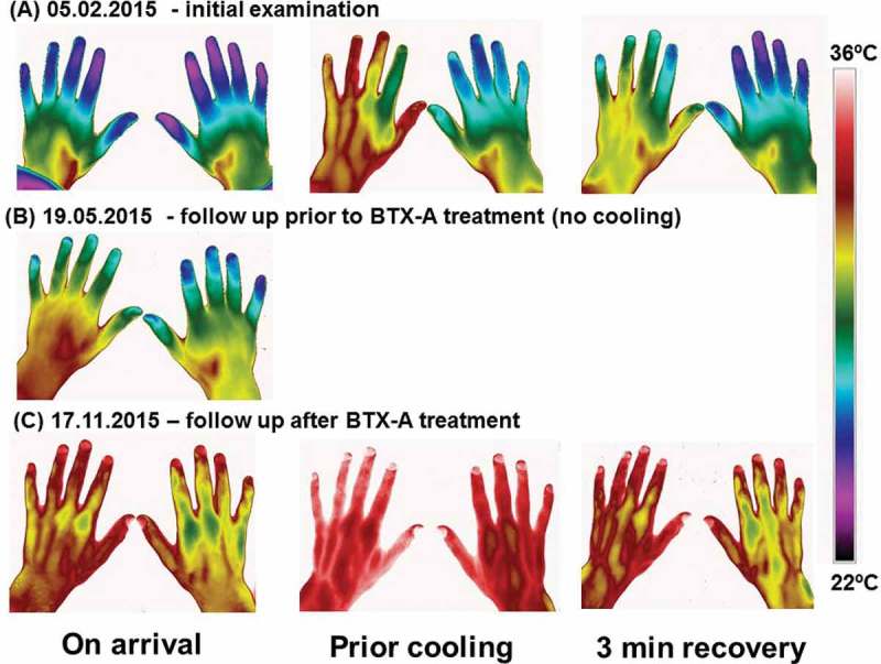
Figure 2.

Thermography before and after treatment. Thermographic images of the hands taken on 3 different occasions. (A) the initial examination 05.02.2015; (B) follow up examination immediately prior to BTX-A treatment 19.05.2015 and (C) follow up examination after BTX-A treatment 17.11.2015. On the first and the third occasion, the patient’s hands was subjected to a mild cold provocation test (2-minute period of convective cooling using a desk top fan). The images were taken after the patient had had sat in the laboratory wearing warm clothes to ensure he was very mildly hyperthermic at the time of the examination. This was confirmed from the patient’s reported subjective feeling as well as from thermal facial images showing that the nose (open arterio-venous anastomoses) was vasodilated. The hand images were taken immediately after the patient arrived at the laboratory (on arrival), after the patient had been warmed up (prior cooling) and 3 minutes after the end of the cold provocation period (3 min recovery).
