
Figure 3.
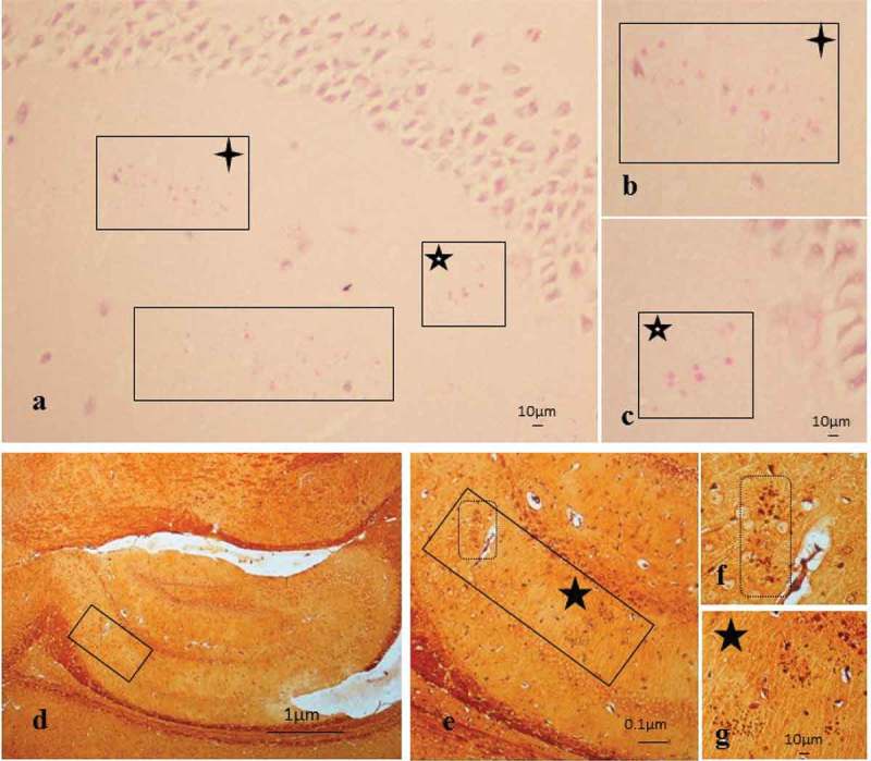
Age-related granules in the brain tissue of infected mice. (a-c) Periodic acid–Schiff reaction in a P. gingivalis tissue section from the hippocampus region showing the mature (large) age-related granules demarcated by boxes. (b-c) Boxes with four-tailed stars in (a and b) and five-tailed star with white core (a and c) are the same regions of the hippocampus, with scale bar shown for clarity. (d) Methenamine silver impregnated tissue section showing an overview of the location of inclusions in the CA1 area of the hippocampus; the same inclusions shown in (e–g) are denoted by boxes and stars.