
Figure 4.
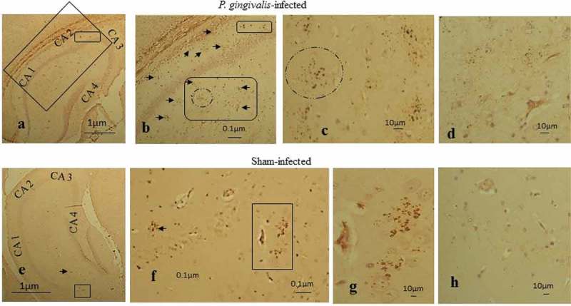
Peptidoglycan immunostaining of infected ApoE−/− mouse brains with hippocampal CA areas labeled (CA1-3). (a-c) P. gingivalis–infected tissue section with numerous clusters located at the boundary of CA2 neurons (smaller box) and (b) in the stratum radiatum CA1 (arrows and larger box with circle). (d) Diffuse immunostaining with anti-peptidoglycan antibody. (e) Fewer age-related granules in the CA1 region of the hippocampus in sham-infected mice indicated by small arrow and box. (f) Arrow pointing to a cluster and two clusters within box and in (g) without the box. (h) Sham-infected ApoE−/− mouse brain devoid of any diffuse staining.