
Figure 6.
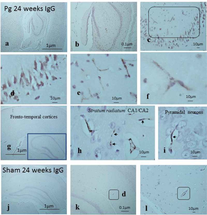
Demonstration of mouse IgG in the cortical brain including the hippocampus to show an impaired BBB due to infection. (a-c) P. gingivalis–infected brain with IgG immunostaining not only on severely damaged capillaries in the stratum radiatum (c, box) of the hippocampus but also on pyramidal neurons. (b-d) Regions from (c) at higher magnification in (e) and (f) (see micron bar). (g-h) Another P. gingivalis–infected brain with IgG immunostaining to illustrate structurally less damaged capillaries in the stratum radiatum (h) of the hippocampus but also (i) not opsonized to pyramidal neurons. (j-l) Sham-infected brains exposed to labeled anti-IgG (k-l) show larger vessels immune-positive for mouse IgG (boxes).