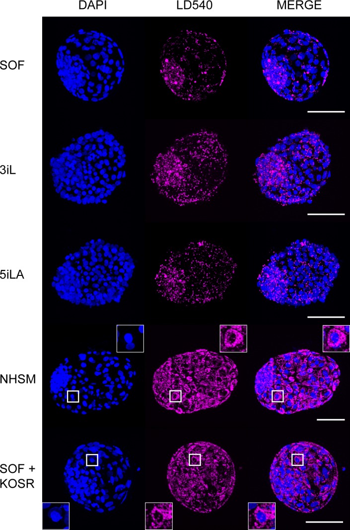
Fig 2. Lipids in bovine embryos.
Representative pictures of embryos cultured in SOF, 3iL, 5iLA, NHSM, and SOF supplemented with KOSR stained by LD540 (pink) for lipid droplets and counterstained with DAPI (blue)are shown. Scale bars represent 100mm. Inserts show a higher magnification of trophectoderm cells with perinuclear lipid droplets.

