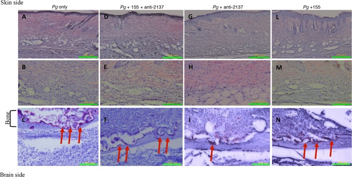
FIG 4.
Histological sections. Shown are representative samples of skin and underlying calvarial bone at the middle of the lesion from each of the following groups: P. gingivalis (Pg) (A to C), combination (P. gingivalis and miR-155 plus anti-miR-2137) (D to F), P. gingivalis and anti-miR-2137 (G to I), and P. gingivalis and miR-155 (L to N). TRAP staining of bone (bottom row, ×200 magnification) and hematoxylin-and-eosin staining of skin (top row, ×100 magnification; middle row, ×200 magnification) are shown. Arrows indicate TRAP-stained multinucleated osteoclasts attached to bone and bone resorption lacunae.

