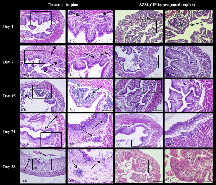
FIG 3.
Histopathology micrographs of bladders derived at weekly intervals from female LACA mice implanted with uncoated or AZM-CIP-impregnated implants and infected with P. aeruginosa PAO1. The tissues were viewed at a magnification of ×40 (a, c, e, g, i, k, m, o, q, and s) and ×100 (b, d, f, h, j, l, n, p, r, and t) under a light microscope. A black box shows the area with or without distinct inflammatory changes in panels a, c, e, g, i, k, m, o, q, and s that are magnified in panels b, d, f, h, j, l, n, p, r, and t, respectively. Arrows indicate inflammatory changes such as the infiltration of neutrophils, and double-headed arrows indicate thickening of the muscularis propria. The bladders from mice in the uncoated implant group exhibit severe inflammation (e and f), massive infiltration of neutrophils (i and j), and ulceration and edema of the lamina propria (e, m, n, q, and r), whereas the bladders from mice in AZM-CIP implant group show initial inflammation and increased vascularity for 1 week, followed by no signs of inflammation. Bars, 50 μm.

