. 2017 Feb 27;6:e20922. doi: 10.7554/eLife.20922

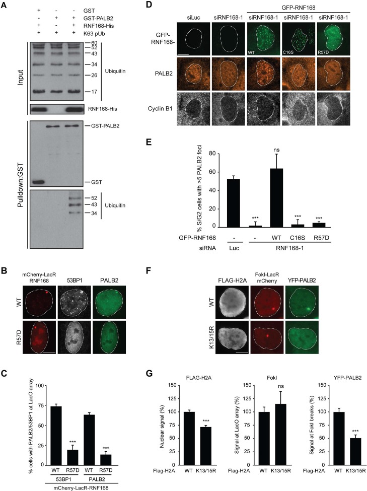
Figure 7. PALB2 associates with K63-polyubiquitin chains via RNF168.

(A) GST pulldowns to assess the binding of GST-PALB2 or GST-alone to K63 polyubiquitin (pUb) chains in the presence or absence of His-RNF168. Blots were probed for GST, ubiquitin and His. (B) Recruitment of 53BP1 (white) and YFP-PALB2 (green) upon tethering of the indicated mCherry-LacR-RNF168 variants (red) to a genomic LacO array in U2OS 2-6-3 cells. Indicated significance is compared to WT for each staining. (C) Quantification of B. (D) PALB2 IRIF formation (orange) in cyclin B1-positive U2OS cells (white) transfected with the indicated siRNAs and siRNA-resistant GFP-tagged RNF168 cDNAs (green). (E) Quantification of D. Indicated significance is compared to siLuc. (F) Recruitment of YFP-PALB2 (green) to DSBs induced by FokI-mCherry-LacR at a LacO array (red) in U2OS 2–6-5 cells expressing the indicated FLAG-H2A constructs (white). (G) Quantification of F. Indicated significance is compared to WT for each staining. Quantified data are represented as mean ± S.E.M. (n = 3), except in E where n = 4. Scale bar = 5 µm.

DOI: http://dx.doi.org/10.7554/eLife.20922.013

Figure 7.

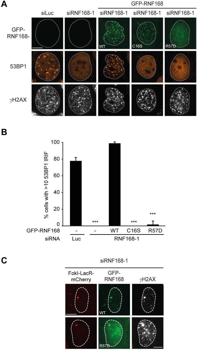
Figure 7—figure supplement 1. RNF168 RING mutants do not support IR-induced 53BP1 focus formation. .

Figure 7—figure supplement 1.

(A) 53BP1 IRIF formation (orange) at 1 hr after 2 Gy in γH2AX-positive cells (white) transfected with the indicated siRNAs and siRNA-resistant GFP-tagged RN168 cDNAs (green) to validate the mutants used in Figure 7D,E. (B) Quantification of A. Indicated significance is compared to siLuc. Quantified data are represented as mean ± S.E.M. (n = 3). Scale bar = 5 µm. (C) Recruitment of the indicated siRNA-resistant GFP-tagged RNF168 cDNAs (green) in RNF168-depleted cells to γH2AX-positive DSBs (white) induced by FokI-mCherry-LacR at a LacO array (red) to validate the R57D mutant used in Figure 7D,E.
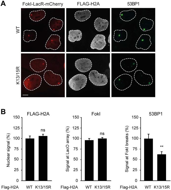
Figure 7—figure supplement 2. Expression of H2A lacking K13 and K15 decreases 53BP1 recruitment to FokI-induced DSBs. .

Figure 7—figure supplement 2.

(A) Recruitment of 53BP1 (green) to DSBs induced by FokI-mCherry-LacR at a LacO array (red) in U2OS 2-6-5 cells expressing the indicated FLAG-H2A constructs (white) to validate the experimental system used in Figure 7F,G. (B) Quantification of A. Indicated significance is compared to WT for each staining. Quantified data represent the mean ± S.E.M. (n = 3). Scale bar = 5 µm.
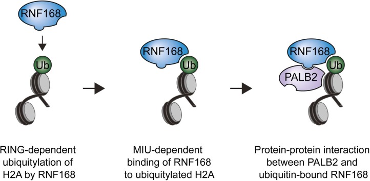
Figure 7—figure supplement 3. Model for RNF168’s role in PALB2 recruitment.

Figure 7—figure supplement 3.

Schematic representation of the RING-dependent ubiquitylation of H2A by RNF168, which is followed by the MIU-dependent binding of RNF168 to ubiquitylated H2A. The direct protein-protein interaction between PALB2 and ubiquitin-bound RNF168 recruits the former to DSBs.