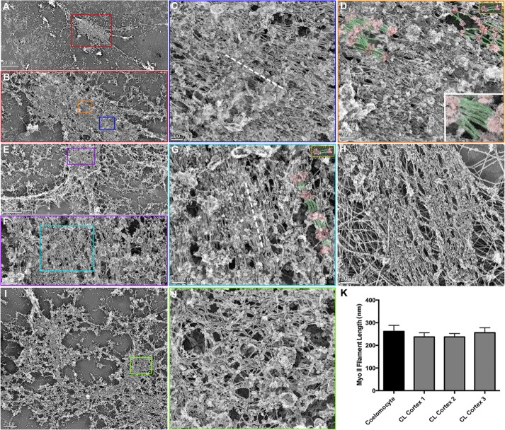
FIGURE 7:
TEM of detergent-extracted, gelsolin-treated cortices reveals tightly packed bundles of myosin II filaments within the CR. Linear arrays of myosin II filaments are evident in detergent-extracted and gelsolin-treated cleavage cortices imaged at low (A, E), medium (B, from red box in A; F, from purple box in E), and high (C, from blue box in B; D, from orange box in B; G, from cyan box in F) magnification. The myosin II filaments are recognized by their characteristic appearance of globular heads and smooth rod/tail regions, their 200–300 nm length (K), and their alignment into head-to-head–associated longitudinal chains. In D and G, a subset of myosin II filament heads have been colored red and tails/rods green, and the gold-bordered insets at the top right show similarly colored isolated myosin II filaments from coelomocytes for comparison. The bottom right inset in D is a high-magnification view of an array of myosin II filaments from a similarly colored cortical CR. The myosin II filaments within the CR region tend to align parallel to the long axis of the CR/cleavage plane (axis indicated by dashed white line in C and G). Myosin II filament organization in the CRs is very similar in appearance to the array of myosin II filaments seen in TEM of gelsolin-treated stress fibers from cultured mammalian LLC-PK1 cells (H; the large filaments in the background are intermediate filaments). In some cortices, myosin II filaments are arrayed in a more isotropic network organization (I, low magnification; J, high magnification from green box in I), which are reminiscent of the patches of myosin II filament networks seen in SIM images of mid-stage cortices (Figures 2 and 3). Scale bars as indicated.

