FIGURE 4:
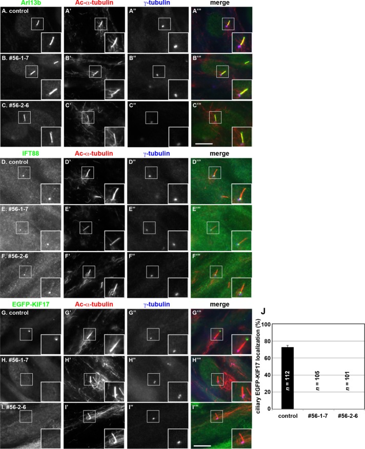
Localization of ciliary proteins in IFT56-KO cells. (A–F) Control RPE1 cells (A, D) and IFT56-KO cell lines 56-1-7 (B, E) and 56-2-6 (C–F) were cultured under serum starvation conditions for 24 h to induce ciliogenesis and triple immunostained for Arl13b (A–C) or IFT88 (D–F), Ac-α-tubulin (A′–F′), and γ-tubulin (A′′–F′′). (G–I) Control RPE1 cells (G) and the IFT56-KO cell lines 56-1-7 (H) and 56-2-6 (I), which stably express EGFP-KIF17 (G–I) and were established by infection of a lentiviral expression vector, were cultured under serum starvation conditions and immunostained for Ac-α-tubulin (G′–I′) and γ-tubulin (G′′–I′′). Merged images are shown in A′′′–I′′′. Insets, enlarged images of the boxed regions. Scale bar, 10 µm. (J) Control, 56-1-7, and 56-2-6 cells positive for ciliary EGFP-KIF17 signals were counted; percentages of positive ciliated cells. Values are means ± SE (error bars) of three independent experiments. In each set of experiments, 50–55 ciliated cells were observed, and the total numbers of ciliated cells observed (n) are shown . Note that no ciliary localization of EGFP-KIF17 was observed in the two IFT56-KO cell lines.

