
Figure 3. The TIRAP R121W allele is loss of function.
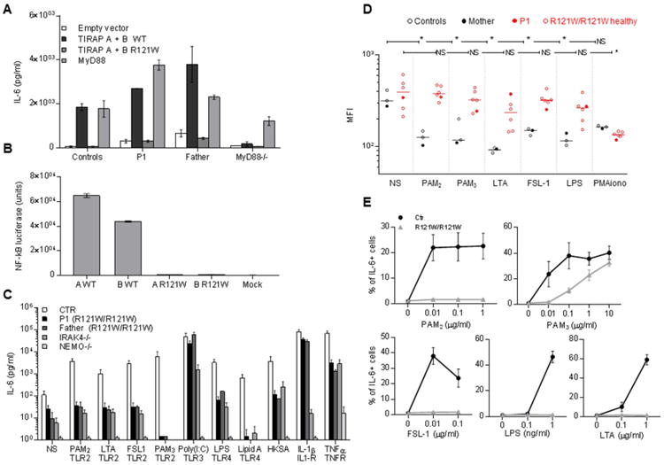
A. IL-6 induction in SV40 fibroblasts from a healthy control, P1, her father, and MyD88-deficient cells, upon transfection with WT or mutant TIRAP isoforms. IL-6 production was measured by ELISA in cells transfected with an empty vector, WT or mutant isoforms A and B, or in MyD88-deficient cells transfected with WT MyD88 (n=3). Error bars show SEM.
B. NF-κB luciferase assay in HEK 293T cells stably expressing a NF-κB-driven luciferase reporter plasmid upon transfection with WT or R121W TIRAP isoform A or B, (n=3). Error bars show SEM.
C. SV40 fibroblasts from healthy controls, P1, her father, an IRAK-4-deficient patient and NEMO-deficient fibroblasts were stimulated with TLR1/2 (PAM3), TLR2/6 (PAM2, purified LTA-SA, FSL-1), TLR3 (poly(I:C)) and TLR4 (LPS and lipid A) ligands, in addition to heat-killed S. aureus (HKSA) IL-1β (IL-1R) and TNF-α (TNF-αR). IL-6 production was assessed by ELISA in supernatants collected 24 h after stimulation and (n=3). Error bars show SEM.
D. Granulocyte L-selectin shedding assessed in several family members (III. 1, IV. 1-5 and V.1) after 1 h of stimulation with PAM2, PAM3, FSL-1, purified LTA, LPS and PMA as an activation control. Mean fluorescence intensity was determined by flow cytometry after the extracellular staining of CD62L. * p values < 0.05 (unpaired t-test) (n=1).
E. PBMCs from several family members (III. 1, IV. 1 to 5 and V.1) were stimulated with PAM2, FSL-1, LTA, PAM3 and LPS for 3 h, subjected to anti-CD3, anti-CD19 and anti-CD14 extracellular staining and IL-6 intracellular staining. The proportion of CD14+ cells positive for IL-6 is shown. 10,000 CD14+ cells were acquired. Each data point represents the mean of all individuals ± SEM.