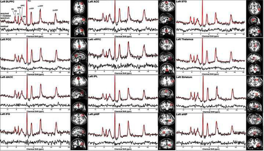
Fig 1.
Voxel placements and representative spectra from each voxel. Only the left sided voxels placements and spectra are shown. Quality of the spectra were high with no significant differences in signal-to-noise (SNR) of PCr between schizophrenia patients and controls. In addition, the Cramer Rao Lower Bound values for both the PC+PE and GPC+GPE were not significantly different between the groups. Legend: DLPFC: Dorsolateral prefrontal cortex; PCC: Posterior Cingulate Cortex; dACC: Dorsal Anterior Cingulate Cortex; IFG: Inferior Frontal Gyrus or Cortex; ACC: Anterior Cingulate Cortex; vPFC: Ventral Prefrontal Cortex; IPL: Inferior Parietal Lobule; pHIP: Dorsal Hippocampus; STG: Superior Temporal Gyrus or Cortex; aHIP: Ventral Hippocampus

