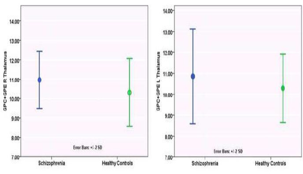
Fig 2.
Comparison of MPL catabolites (GPC+GPE) in the thalamus between schizophrenia patients and healthy controls. In addition, the dorsolateral prefrontal cortex, ventral hippocampus, inferior frontal cortex, inferior parietal lobule and superior temporal gyrus also showed diagnosis main effect (Overall model, F(24, 614)=8.19, p<0.001). While GPC+GPE levels were elevated in bilateral thalamus and right ventral hippocampus it was reduced in the right dorsolateral prefrontal cortex, right inferior frontal cortex, and bilateral inferior frontal lobule, superior temporal gyrus in schizophrenia compared to controls. See Table 2 for percentage differences at these voxels-of-interest between the groups.

