
Fig. 7. Re-expression of GSK-3β inhibits LPA-induced cell proliferation.
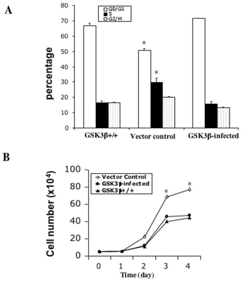
GSK-3β−/− cells were infected with the HA-GSK-3β retro-virus or the control virus as detailed under “Experimental Procedures.” GFP-positive cells from HA-GSK-3β retrovirus-infected cells (GSK-3β infected) and control virus-infected cells (Vector control) were sorted, replated, and cultured for growth response to LPA. A, cell cycle analysis of the GSK-3β-infected, vector control, and GSK-3β+/+ cells. The cells were starved for 16 h and incubated with 10 μM LPA for 24 h before collection by trypsinization. Cells were stained with propidium iodide for flow cytometric analysis of cell cycle progression. B, growth curves of the GSK-3β-infected, vector control, and GSK-3β+/+ cells. Cells were seeded in 6-well plates (5 × 10⁴ cells/well). After attachment, cells were starved for 16 h and cultured in serum-free medium containing 10 μM LPA. Cell numbers were determined at 24-h intervals by counting in a hemocytometer. For both panel A and panel B data are presented as means ± S.D. of duplicate assays from three independent experiments. Statistical differences between GSK-3β-infected cells and vector control cells are indicated by an asterisk (p < 0.01).