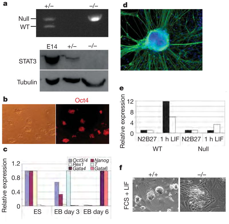
Figure 3. ES-cell propagation in 3i does not involve STAT3.
a, Top: genomic PCR for null and wild-type (WT) Stat3 alleles in heterozygous and Stat3 homozygous null ES cells. Bottom: immunoblot analysis of heterozygous and Stat3 homozygous null ES cells. E14, E74Tg2a ES cells. b, Oct4 immunostaining of Stat3-null ES cells. c, Quantitative RT–PCR analysis of undifferentiated Stat3-null ES cells and derivative embryoid bodies (EB) at days 3 and 6. d, Stat3-null ES cells generate morphologically differentiated cells expressing the neuronal marker βIII-tubulin (TuJ1 immunoreactive). e, Quantitative RT–PCR analysis of Socs3 (Stat3 target gene; filled columns) and Egr1 (ERK target gene; open columns) expression in undifferentiated Stat3 wild-type and null ES cells grown in N2B27 alone or stimulated with LIF for 1 h. f, Stat3-null MF1 ES cells differentiate in the presence of LIF and feeders, in contrast with wild-type MF1 ES cells, which remain undifferentiated.

