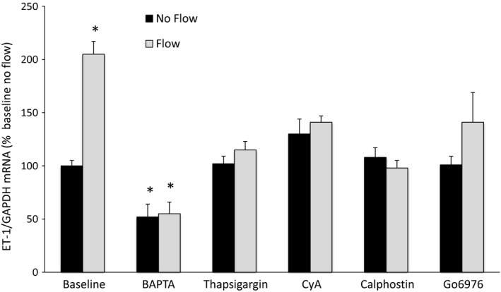
Figure 1.

Role of Ca2+ signaling pathways in flow‐stimulated endothelin‐1 (ET‐1) mRNA levels in MPK‐CCD cells. Cells were preincubated with vehicle, 50 μmol/L BAPTA‐AM, 200 nmol/L thapsigargin, 3 μg/mL cyclosporine (CyA), 0.1 μmol/L calphostin C, or 200 nmol/L Go6976 for 30 min, followed by exposure to static conditions or shear stress at 2 dyne/cm2 for 2 h in the presence of the same agents, then determination of ET‐1/GAPDH mRNA levels. N = 8–12 each data point. *P < 0.05 versus baseline (vehicle alone) no flow conditions.
