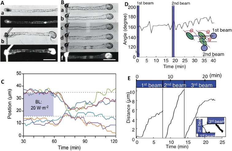
Figure 3.
Chloroplast behavior during movement. A. Two-celled protonema of Adiantum capillus-veneris. (a) Protonema with a short tip cell and a long basal cell (top), with a DAPI-stained image (below). (b) Protonema enucleated by ligation and separation of the nuclear region (top), with a DAPI-stained image (below). Bar: 100 µm. B. Chloroplast movement in an enucleated cell. (a) Enucleated cell. (b) Chloroplast movement induced by polarized blue light. Chloroplasts are on both sides of the cell. (c) Partial cell irradiation with a weak blue microbeam. (d) Induced accumulation response. (e) Avoidance response induced with a strong microbeam. (f) A lack of a nucleus in the cell was confirmed by DAPI staining. Bar: 100 µm. C. Signal lifetimes for avoidance and accumulation responses were calculated in a prothallial cell. Chloroplasts outside of the strong blue light beam migrated into the former beam-irradiated area following a lag period (i.e., duration of the avoidance response) after the light was switched off. They then started to disperse (i.e., dark positioning) after a lag period (i.e., duration of the accumulation response). Each chloroplast movement is indicated by different colored line. D. Chloroplasts change directions without rotating during the accumulation response. The angle between the long axis of chloroplasts and the direction toward the first beam was continuously measured. Even though the destination of chloroplasts changed to the second beam, the angle did not change. The inset presents a diagram of the experimental procedure. The data were obtained from Movie 1. E. Avoidance responses induced when half of a chloroplast was irradiated three times with strong and continuous blue light. The data were obtained from Movie 3. The avoidance response occurred after a short lag period. The inset presents the chloroplast movement path. Figures A and B are reproduced from Wada 1988,81) C is from Higa and Wada 2015,76) D and E are from Tsuboi et al. 200975) and Tsuboi and Wada 201171) with copyright permission from Japanese Society of Plant Physiologists, Wiley, Botanical Society of Japan, respectively.

