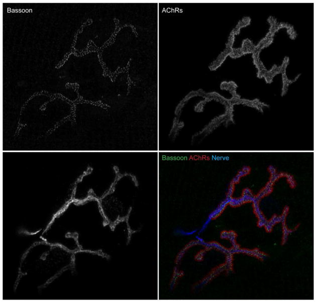
Figure 4.
Bassoon localization at an NMJ. Immunohistochemistry detection of active zone specific protein Bassoon in an NMJ of an adult (4 month old) wild-type C57Bl/6 mouse. Longitudinal sections of gastrocnemius muscle were stained with primary antibodies against Bassoon (green), SV-2 and neurofilament to visualize nerve terminal (Nerve, blue), and Alexa Fluor 594-labeled α-bungarotoxin to visualize acetylcholine receptors (AChRs, red). An NMJ was imaged on a confocal microscope and deconvolved. Scale bar represents 5 μm.

